Skin.
Our interface with the world. When something goes wrong with skin, people notice. Scars, acne, a change in pigment. Wounds that refuse to heal and chronic conditions like psoriasis. When skin doesn’t behave properly, it hurts.
For over 25 years, molecular biologist Jonathan Jones has been looking for ways to help speed the epidermal healing process. As a child in Wales, he’d suffered from itchy red patches of eczema, an annoying condition that eventually got him thinking about skin in a scientific way. Recently, that interest paid off with the surprising discovery that skin cells “walk” during wound healing. The finding could provide new treatment options for injuries, skin cancer, and other disorders.
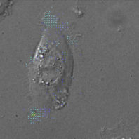
Now a professor and director of the Washington State University School of Molecular Biosciences, Jones showed that individual skin cells called keratinocytes move step-by-step to bridge and repair damaged tissue.
Skin is a multilayered organ and keratinocytes form the protective outer layer. Further down lie the basal cells, some of which “button” the skin to underlying connective tissue by tiny structures called spot wells. In 1991, Jones began studying the proteins in spot wells as a way to improve skin grafting for burn victims. The idea was to coat a burn site with the proteins before placing the graft in hopes that it would attach more effectively.
“What I didn’t realize was that those proteins also play a role in the migration of skin cells,” he says. “The finding was unexpected as we thought spot wells were only good for holding skin to connective tissue … that they’d actually inhibit cell movement.”
The revelation refocused Jones’ efforts. Since 2006, he has painstakingly dissected spot well proteins to pinpoint their exact method of action. The work promised to explain how skin cells move both during wound healing and in the spread of squamous cell carcinoma, a form of skin cancer.
The breakthrough came recently when Jones, postdoctoral research associate Sho Hiroyasu, and graduate student Zachary Colburn found that the proteins act like quarterbacks, deciding which end of the keratinocyte will be the front and which the rear. They then generate signals telling the cell to “walk” from point A to point B.
The proteins do this by activating muscle-like forces in the keratinocyte necessary to march across an abrasion or laceration. During this process, Jones says spot well proteins temporarily release the cell from the underlying connective tissue, allowing it to “lay” new skin over the wound.
The system tends to break down in diabetics and the aged, he says. “Their skin cells don’t move very efficiently over the wound surface so they develop chronic ulcers which don’t heal properly. Secondary infections contribute to the poor healing.”
In time, Jones hopes to develop ways to enhance the activity of spot well proteins to promote faster, more effective healing for all types of wounds, including grafts.
“The irony is the same proteins also promote migration of tumor cells,” he says. Skin cancer cells have co-opted the same mechanism to migrate through skin and to other parts of the body.
“So, it’s a bit of a two-edged sword. There is a lot of interest in these mechanisms for blocking the spread of tumors but you don’t want to stimulate them too much or skin cells go mad. And, if you inhibit them too much, it prevents normal wound healing.”
While Jones’s findings came through the study of individual skin cells, in the body they move as collective sheets of millions. His laboratory is now using 3D modeling to better understand how these sheets behave during skin repair. So far, it appears leader cells at the front edge of a wound express the proteins necessary to get the whole sheet moving to repair itself.
The potential applications for mending skin and other tissues have Jones and his colleagues excited. Already Colburn is investigating lung diseases and the role of spot well proteins in recovery from pulmonary injury and infections like influenza.