Despite its many mysteries, biomechanics serves up surprises about strained muscles and bones broken and mended.
Earlier this year, at the ripe age of 38, Bernard “Kip” Lagat ’01 became the fastest American ever to run two miles indoors. It was a feat of both speed and longevity, helped in large part by a fluid, seemingly effortless running form the New Yorker describes as “perfect.”
It was not always so. In fact, Lagat’s performance, as well as two Olympic medals and several other American records, may never have taken place without the long tutelage of James Li MS ’87 MS, ’93 PhD, who recruited Lagat from Kenya’s Rift Valley Province in the mid-’90s.
“He was pretty good,” Li recalls “but I would venture to say that he was not as smooth as he is now.”
(Photo Robert Hubner)
Li, who continues coaching Lagat while serving as a coach at the University of Arizona, has a résumé that includes more than ten years with WSU’s track and field and cross-country programs, as well as the collegiate 800-meter title in his native China.
It also helps that his WSU master’s degree is in biomechanics.
Biomechanists, as they are sometimes called, straddle the worlds of engineering and biology. Like engineers, they study the physics of objects in motion or under strain, but their objects are living things. That introduces a host of complications. An engineer can design a vehicle on the known properties of steel, wheels, a motor, and so forth. A biomechanist will wrestle with muscle cells of varying power and body parts articulating under the direction of their owner’s neurology, not to mention personal style.
Seemingly simple questions quickly get complex, like, “How do we hold up our head?”
“Scientists don’t really understand that,” says Anita Vasavada, an associate professor of bioengineering and neuroscience who has made one of the most sophisticated models of neck musculature.
David Lin, a biomedical engineer and Vasavada’s husband, is currently struggling to model how a human trips and falls, a sort of Lagat gone bad. It’s a complicated process, with lots of parts—arms, legs, a torso—moving in three dimensions and sometimes acting against each other. For simplicity’s sake, his model has no spine or arms.
“You’ve got to pick your battles,” he says, “and you’ve got to make a hypothesis about what you think is important and then you create a model that provides a representation of whatever that is. Then you run your model and try to make some conclusions.”
In spite of the challenges, biomechanists are indeed managing to draw conclusions, or at least some striking intermediary insights.
In a Moscow, Idaho, symposium earlier this year, WSU biomechanists discussed with other Northwest researchers the pigeon-toed running style of grizzly bears; ways accelerometers can detect dyskinesia, a side effect of Parkinson’s disease treatment; and how a concussion might affect the way an adolescent walks.
On the Pullman campus, Vasavada is helping decipher the muscular mechanics of whiplash, gender differences in neck-pain sufferers, and potential pitfalls in how tablet owners interact with their screens. Other researchers are working to make a safer softball by seeing in excruciating digital detail how one can break bones and other body parts. Nearby, other researchers are finding ways to repair broken bones by getting the body to heal in synch with synthetic compounds produced on a 3D printer.
Few body parts are called upon to do as much as the human neck.
A curved, narrow post, it has to hold up a head that weighs more than a gallon of milk and keep it stable enough for consistent vision and hearing. At the same time, it needs to make large movements, like looking over your shoulder.
It does all these things quite well, but its dual purposes make it inherently flawed.
“You have these conflicting demands of mobility and stability,” says Vasavada. “When you have too much or too little of those, most likely you’re going to have pain.”
Vasavada came to study the neck by way of the leg, which had been extensively modeled by her Northwestern University doctoral advisor, Scott Delp. His model helped analyze problems like the crouch gait that has children with cerebral palsy walking with excessively bent knees. His work pointed the way for surgeries to remedy the problem. Vasavada came to Delp after several years working with cadavers and implants in a spinal biomechanics lab, so he suggested she try modeling the neck.
She ended up developing the first musculoskeletal head and neck model based on the neck’s actual anatomy. With 20 color-coded muscles in play, it looks like a scaffold of multicolored pick-up sticks set on their ends and running at odd angles among the shoulders, spine, chin, and head.
She has since used her model in an extensive study of whiplash. It is the most common motor vehicle injury, as well as the most poorly understood. This is largely because the neck offers plenty of parts to be injured, with three joints on each of its seven vertebrae, as well as ligaments, discs, nerves, and arteries. Focusing on the neck muscles, Vasavada collaborated with forensic engineers who had volunteers sit in a car seat and experience a five-mile-an-hour rear-end collision. High-speed video documented their body movements, while electrodes recorded their muscle reactions.
As Vasavada replays the video in her McCoy Hall lab, it’s easy to see a participant’s head snap backwards as the collision thrusts his body forward. Earlier research tended to focus on this motion and the strain it places on the large sternocleidomastoid muscles that run from the collar bone toward the ears.
“Most people have focused on the early phase of whiplash and the sternocleidomastoid,” says Vasavada, “but most of the pain that people report is on the backside of the neck.”
The video suggests why as the crash victim’s head rebounds and shoots forward. Vasavada put these movements into her neck model, which could then calculate the forces and potential strains on other neck muscles.
Muscles are generally injured when they are lengthened too far while tensed. “It’s called an eccentric contraction,” says Vasavada, explaining that it releases chemicals like creatine kinase, which lab tests can use as a measure of muscle breakdown. It can also rupture muscle cell membranes.
Vasavada’s analysis showed significant lengthening of the sternocleidomastoid. It also showed that the strain rate was higher in the muscles behind the neck, possibly explaining the soreness that whiplash victims report there.
Vasavada has also tackled another neck enigma: The inordinate percentage of women who experience neck pain.
Estimates vary, but women are as much as three times more likely than men to experience chronic pain after a whiplash injury. There could be cultural reasons, says Vasavada. They could drive smaller cars that absorb less of a collision’s energy. They could more often be passengers and less aware of an impending rear-end collision. They could have different reactions to pain or a higher threshold for seeking medical attention.
Or it could be biomechanical.
With that in mind, Vasavada measured the neck length and neck strength of 90 subjects and found 14 pairs of men and women with heights and neck lengths within half a centimeter of each other. The women ended up having heads that were only slightly smaller in circumference—about 3 percent—than their male counterparts. But their necks were on average 16 percent smaller. In other words, their small necks were being made to work 33 percent harder than the thicker necks of their male counterparts.
“They’re kind of closer to their limit and possibly more likely to fatigue just by the simple act of holding up their head,” says Vasavada, “much less than when you put them in the Army and put a heavy helmet on them, those kinds of things.”
Most recently, Vasavada and Lin have been pondering how all our necks will fare in the rapidly dawning age of the tablet computer.
In some ways, users interact with them much as they have with old-school technology like books and newspapers, says Vasavada.
But newspaper readers might move more, reducing fatigue, she says. Also, “It may be that tablets force you to stay in that same position, you’re just so enthralled. Especially with games, and that’s where people have the great potential to get neck pain.”
“People report being on their iPad for hours, six hours a day,” adds Lin. “Most of us aren’t reading a book six hours a day.”
Vasavada and Lin photographed subjects using iPads in a variety of postures: with the iPad flat on a table, on a stand, on a user’s lap. Because necks vary from person to person, with the actual positions of vertebrae hidden by muscles and other tissues, they also took x-rays.
Sure enough, they saw the tablet user’s heads move forward, activating more neck muscles to hold up and balance their 10-pound heads.
“When you’re in this head-forward posture,” says Vasavada, “your muscles need to be anywhere between two and a half to three times more active.”
Over the years, scientists and regulators have determined the best ergonomics for desktop computers. But despite their growing popularity says Vasavada, “There are no guidelines for tablet PCs at this point.”
“So that’s why these studies are important,” says Lin.
Lloyd Smith is one of the nation’s leading experts on bats and balls and what happens when they collide. His Sports Science Laboratory has tested bats for the National Collegiate Athletic Association and blown away some of baseball’s storied assumptions, like the myths that a corked bat hits the ball farther and that baseballs today are livelier than, say, the late ’70s.
Modeling the properties of bats was easy, he says. Modeling the properties of a ball, less so. And things really get tough when you introduce the human element and ask how to improve player safety.
“There’s a number of questions you can ask,” he says one afternoon. “One is: If you get hit by a ball, when does injury occur? What is being injured? Are you breaking bone? Are you bruising? Are you causing internal injury? And if you have that injury, what are the criteria for when something is injured? Is it based on acceleration? Is it based on rate of deformation? Is it based on force or stress? Even trying to figure out what the right injury criteria are is a challenge and not something that people know.”
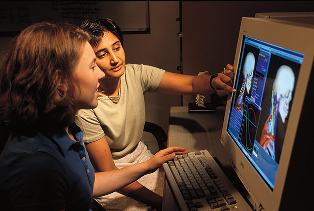
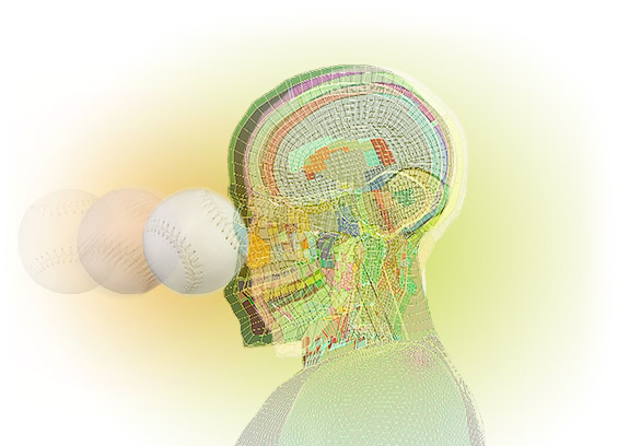
To get started, Smith has called on Derek Nevins ’10 BS, ’12 MS Engineering. A former Vasavada graduate student and now a research project engineer, he has repeatedly slammed a ball into a virtual human head.
The head comes from the Total Human Model for Safety developed by the Toyota Motor Corporation and Toyota Central R&D Labs. Based on data drawn from cadavers and high-resolution CT scans, the color-coded model gives a detailed look at the properties of not only an adult human head as a whole, but components like teeth, eyeballs, skin, skull, meninges, even different sections of the brain.
“Play this model so he can see the brain slosh around,” Smith says to Nevins. “I’m always amazed when I watch this.”
Nevins raps his keyboard and soon a softball is careening in slow motion toward the forehead. The ball compresses as the head lurches back, then forward. Indeed, the brain moves slightly behind the head, sloshing ever so slightly inside the cranium.
In some ways, the video is largely for scientific street cred.
“Many of the people we work with have no idea what we’re doing,” says Smith. “We’re working with softball coaches and baseball coaches. So actually the video for them is very helpful because they believe very little of what we do, being scientists. Having something that looks realistic to them says, ‘Wow, OK, I can believe that.’”
Moreover, says, Nevins: “You see that the shock wave propagates through those different materials differently. You get a small deflection of the bone but then you can see the brain sort of jiggle inside the skull. With that jiggling, there’s that oscillatory behavior that might lead to peak strains in a place that you wouldn’t necessarily have anticipated. But we can investigate that with the model here.”
Some answers will lie outside the science. For example, the researchers could find a safer ball, and a rule-setting federation could adopt it, but it may be so lacking in liveliness that it’s closer to a beanbag than a baseball.
“If the ball speed is 25 miles per hour, is that a game that anybody is interested in playing?” says Smith.
For now, the model is helping answer one of the lab’s first and easiest biomechanical questions—When does bone break?—by providing bone stresses that the researchers can compare with known stresses from actual cadaver and impact studies. They can also see the stress contours of balls with different stiffnesses as they hit the forehead.
Already, the work has shown that two outwardly identical softballs, each approved for the same level of play, can vary so much that one can have a 63 percent greater impact on a head.
Their work has also yielded a striking, counterintuitive revelation: If you are going to get hit in the head by a ball, you may be better off if you don’t see it coming.
A fielder caught unawares is more likely to be facing the ball. He or she might lose some teeth or break a nose, or the ball might hit the stiff forehead, in which case a softer ball will cause less damage.
But a player who sees the ball coming might turn his or her head, exposing the softer temporal area. Ray Chapman, the only professional baseball player killed by a pitched ball, was hit in the temple.
“The injury that is most severe, a temporal impact, really isn’t affected by the stiffness of the ball,” says Smith. “So for a federation, if their goal is to reduce injury, OK, then lower the stiffness of the ball and you reduce injury in this case. If your goal is to reduce fatalities, well, reducing ball stiffness isn’t going to help.”
Ten years ago, Washington State Magazine had a cover story on the work of engineering professors Amit Bandyopadhyay and Susmita Bose. The cover had a picture of their son Shohom and the headline, “It’s not easy to mimic nature.”
It remains a recurring theme of their work, if not biomechanics in general, but they have been having remarkable success nonetheless.
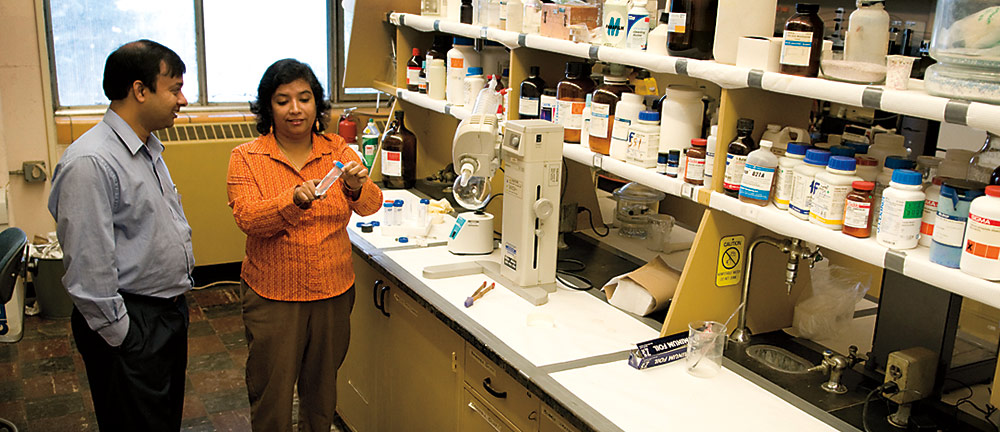
Since meeting at Rutgers University in the late-’90s, they have spent much of their careers creating a synthetic bone, with Bandyopadhyay focusing on its engineering and Bose on the chemistry and application to human health. At first glance, it might seem easy. Bone is mostly calcium phosphate. Shape it, bake it into a ceramic, and you’re good to go.
But chemistry, physics, and biology, particularly at the cellular level, soon get in the way.
“There are many issues,” says Bandyopadhyay. “It’s really fishing in a big ocean.”
Ideally, a physician can tailor some replacement bone to the size and shape of a break and insert it into the body. The body will then use the replacement as a scaffold on which it will build new bone as the replacement dissolves. Throughout the process, the replacement will perform like regular bone, providing structural support until its natural replacement takes over.
If the replacement dissolves too fast, the bone breaks again. If it doesn’t dissolve, a patient can get too much new bone, even cancer.
“It can actually kill a patient,” says Bose.
One challenge is that bone has, in addition to calcium phosphate, trace elements whose function can be largely a mystery. By adding just half a percent of strontium oxide, which is already in use as a drug to treat osteoporosis, an OK bone material becomes exceptional. Similarly, Bose and Bandyopadhyay have found the addition of silicon and zinc more than doubles the fake bone’s strength.
Another challenge: Depending on where they are placed, different materials heal at different rates. Healing that takes three to six months in a jawbone can take nine to 12 months in the spine.
All while patients tend to want quick results.
Basically, says Bandyopadhyay, Mother Nature has 10 to 20 years from a baby’s birth to grow and mature a bone.
“However,” he says, “when there’s a bone fracture, you cannot tell a patient, ‘I’ll put in something and it’s going to be healed in 20 years.’ The patient wants the fracture to be healed if possible in six days … Essentially we need to learn from what Mother Nature has done but we also need to learn how we can accelerate the process so things happen faster than the natural kinetics.”
In spite of all these challenges, they’re starting to make it work. Two years ago, the couple drew national attention when, with the help of a $1.5 million National Institutes of Health grant and equipment support from the M.J. Murdock Charitable Trust, they produced their bone-like material on a 3D printer. The work bolstered the possibility that doctors will be able to custom order replacement bone tissue in a few years. In vitro lab tests showed the material was biologically compatible, as have later tests involving rats and rabbits.
“So far the compositions we have done in the animal model show significant promise in terms of bone formation in the scaffold as well as some blood vessel formation,” says Bose.
That said, it’s still hard, she says.
“We will always keep doing as scientists or engineers the research that needs to be done to solve the problems related to human health,” she says, “but it will still be difficult to mimic Mother Nature.”
The work of WSU’s current biomechanists is a world removed from James Li’s master’s studies in the mid-’80s, when he had none of the high-speed video and computing power behind today’s biomechanical models. Using 35-millimeter film, he analyzed steeplechase hurdlers by focusing on a dot on the hinges of their moving joints.
A decade later, he started videotaping Bernard Lagat and analyzing his form. He relied on observation, not scientific measurments, but he could detect several flaws that on the track translated into crucial seconds: a high knee lift that would affect the landing angle of the foot, a long hang time that suggested too much energy going up, not forward.
Li suggested changes so small and subtle, they might not have yielded any significant data if he could have measured them. As it was, he couldn’t.
Still, he says: “A biomechanics education, the background there, was extremely important, because it gave me the basic principles, the basic science of it, when you have Newton’s laws and angular forces, torque, momentum, and all those things.”
For his part, Lagat earlier this year told Track and Field News that biomechanics has prolonged his career.
“[If] I could credit one person with that, it’s my coach,” Lagat said, recalling videotaped sessions on a treadmill and repeated suggestions to tweak his technique.
“I wasn’t the guy who knew how to run really good like that,” Lagat said. “Coach Li was the one, back in Pullman.”
Li is reluctant to take credit for Lagat’s longevity. But he does say that, now, Lagat’s form is, “efficient, light—it’s like he’s flowing. The flow is just very smooth and he’s known in the track community as probably the most smooth runner.”
Thanks in part to biomechanics, Lagat is one of the world’s great bodies in motion.
Show me where it hurts: To begin calculating the forces of a ball on a human head, WSU’s Sports Science Laboratory turned to the virtual head of Toyota’s Total Human Model for Safety. Based on data drawn from cadavers and high-resolution CT scans, the color-coded model helped researchers determine that two balls approved for regulation play can have vastly different effects on a head. They also saw how where the ball hits can make a huge difference in its potential for injury and death. Staff illustration